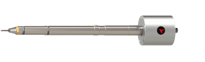
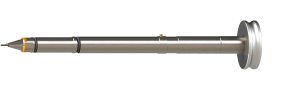
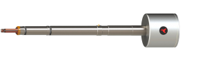
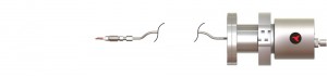
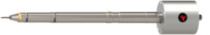
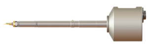
Our specimen holders extend the capabilities of the Transmission Electron Microscope. Conduct your experiments in-situ in the TEM.
Our sample stages extend the capabilities of the Scanning Electron Microscope / Focused Ion Beam Microscope. Conduct experiments inside the SEM or prepare your specimen inside the FIB.
To get the most out of your TEM holders use Hummingbird Scientific's selected TEM holder accessories.
With a custom TEM sample holder from Hummingbird Scientific, Nielsen et al. were able to observe, in situ, crystal nucleation and growth of calcium carbonate (CaCO3) in solution at the nanometer scale. The careful design of the cell and sample holder gave them control over the reaction environment which allowed the researchers to directly investigate nucleation. You can watch the attachment of an aggregate and subsequent transformation into crystal by clicking the image above.
[Read More...]
Reference:
Nielsen MH et al. Structural evolution, formation pathways and energetic controls during template-directed nucleation of CaCO3. Faraday Discuss. 2012; 159:105-121. Image copyright 2012, Royal Society of Chemistry.
Researchers at UC Davis, Lawrence Livermore National Laboratory and Pacific Northwest National Laboratory worked with a continuous flow in-situ liquid stage developed by Hummingbird Scientific to obtain electron energy loss spectroscopy (EELS) spectra from nano-particles in colloidal suspension using aberration-corrected scanning transmission electron microscopy (STEM).
[Read More...]
References:1) Jungjohann, Katherine L., James E. Evans, Jeffery A. Aguiar, Ilke Arslan, and Nigel D. Browning. "Atomic-Scale Imaging and Spectroscopy for In Situ Liquid Scanning Transmission Electron Microscopy." Microscopy and Microanalysis 18, no. 03 (May 2, 2012): 621-627. ABSTRACT Paper and images copyright © 2012, Microscopy and Microanalysis.
2) R.R. Unocic, L. Baggetto, K.A. Unocic, G.M. Veith, N.J. Dudney, and K.L. More. "Coupling EELS/EFTEM Imaging with Environmental Fluid Cell Microscopy." Microscopy and Microanalysis 18 (Suppl 2), 2012 1104-1105