
SEM Liquid Flow
Available For:




.jpg)


Key Features and Capabilities
Reproducible Screw-Free Liquid-Cell Assembly
Removable, Swappable Tip and Tubing
Flexible Liquid Cell Operation
Multimodal Imaging
Optimized for EDS
Integrated Liquid Heating
Dual-Flow Liquid Mixing
Biologic SP-200 Potentiostat
Built for Diverse Applications


Featured Research
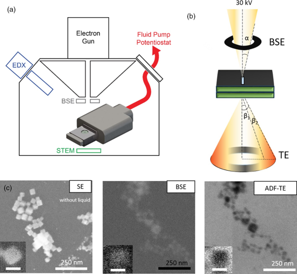
Dynamic multi-detector liquid-phase SEM imaging of nanoparticles in solution
a) Schematic of LC-SEM setup with BSE, STEM, and EDX detectors. b) The electrons scattered from the liquid cell are collected in both forward and backward directions. The forward scattered electrons are collected by the STEM detector, and the backward scattered electrons are collected by the BSE detector. c) Cu2ONPs imaged with the different electron detectors: SE, BSE, and ADF-TE from left to right. The BSE and ADF-TE images were acquired with 0.1 M KHCO3 electrolyte inside the assembled liquid cell. Scale bar is 25 nm in the inset.
Reference: Aram Yoon, et al. Microscopy and Microanalysis (2021). DOI:10.1017/S1431927620024769
Copyright © Microscopy Society of America 2021. Published by Cambridge University Press.
Selected Publications
Calvin Parkin, Tyler Mefford, Jongwoo Lim, Aram Yoon, Rui Serra-Maia, Pawan Kumar, See Wee Chee, William C. Chueh, Daan Hein Alsem, Norman Salmon, Eric A. Stach. “In-Situ Liquid-Electrochemical X-Ray and Electron Microscopy for Multi-Modal Energy Materials Characterization”. ECS Meeting Abstracts (2024)
Alejandra Londono-Calderon, Srikanth Nayak, Curtis L. Mosher, Surya K. Mallapragada, Tanya Prozorov. “New approach to electron microscopy imaging of gel nanocomposites in situ”. Micron (2019)
Alejandra Londono-Calderon, Wan Sun, Zengyi Shao, Daan Hein Alsem, Tanya Prozorov. “Correlative Microbially-Assisted Imaging of Cellulose Deconstruction with Electron Microscopy”. Microscopy and Microanalysis (2018)
D.A. Fischer, D.H. Alsem, B. Simon, T. Prozorov, N. Salmon. “Development of an Integrated Platform for Cross-Correlative Imaging of Biological Specimens in Liquid using Light and Electron Microscopies”. Microscopy and Microanalysis (2013)