
How do structure and dynamics shape the behavior of biological and soft materials?
Hummingbird Scientific in-situ sample holders enable real-time imaging of biological and soft materials in hydrated environments, revealing structural changes, particle interactions, diffusion, and responses to controlled liquid conditions. Conduct multimodal TEM, SEM, and X-ray experiments using continuous-flow and mixing configurations suited to the workflow.
Each holder is designed for reliable performance, reproducibility, and ease of use. Explore relevant experiment types, published examples, and compatible Hummingbird products below.

Which type of experiment best matches your research?

Biomaterial induced corrosion
Visualize biomaterial-induced corrosion processes and nanoscale structural evolution in real time under liquid environments.

Biomaterial induced corrosion
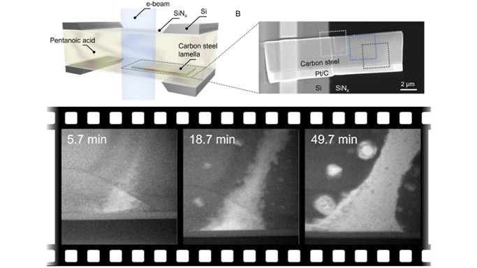
Real-time nanoscale mapping of pentanoic acid–induced corrosion in carbon steel
In-situ imaging revealed how biofeedstock-derived acids drive corrosion in carbon steel. Correlative analysis linked microstructure to initiation and propagation.
- Liquid-phase TEM of steel lamellae in pentanoic acid, combined with TKD, EDX, and thickness mapping.
- Strain-driven corrosion observed with accelerated localized attack and minimal grain boundary activity.
- These results highlight liquid-phase TEM for probing biomaterial processing environments, enabling predictive, real-time evaluation of material stability in complex organic media.
Reference: Zhiheng Lyu, et al, ACS Nano 19, 31, 28315-28325 (2025) DOI: 10.1021/acsnano.5c06142
Image copyright © 2025 American Chemical Society

Biomolecule-templated growth
Capture biomolecule-templated mineral growth and self-assembly while directly observing nanoscale structural evolution.
Biomolecule-templated growth
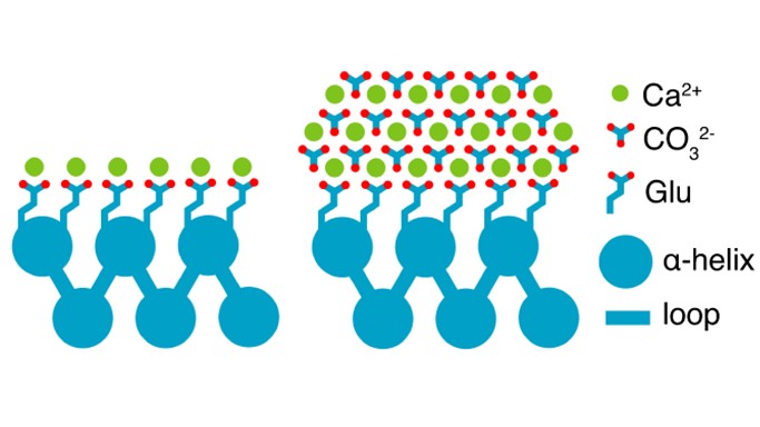
Uncovering the dynamics of protein-engineered nano-calcite nucleation and assembly
De-novo helical repeat (DHR) proteins were used as programmable templates to direct controlled calcium carbonate nucleation, growth, and assembly, captured in real time via in situ liquid-phase TEM.
- DHR monomers and protein–Ca2+ assemblies were observed directly, with engineered α-helical motifs pre-organizing Ca2+ ions to mimic calcite coordination.
- Nano-calcite nucleated with non-natural {110}/{202} facets, followed by nanoparticle attachment driving oriented mesocrystal assembly with tunable size and polymorph.
- The results establish in-situ liquid phase TEM as a powerful platform for decoding and programming biomolecule-templated mineralization, enabling design of hybrid materials for carbon sequestration, catalysis, and bioinspired systems.
Reference: Fatima A. Davila-Hernandez, et al, Nat. Commun. 14, 8191 (2023). DOI: 10.1038/s41467-023-43608-1
Image copyright © 2024 Springer Nature Limited

Biomineralization processes
Visualize nucleation, growth, and phase evolution during biomineralization under liquid-phase conditions.
Biomineralization processes
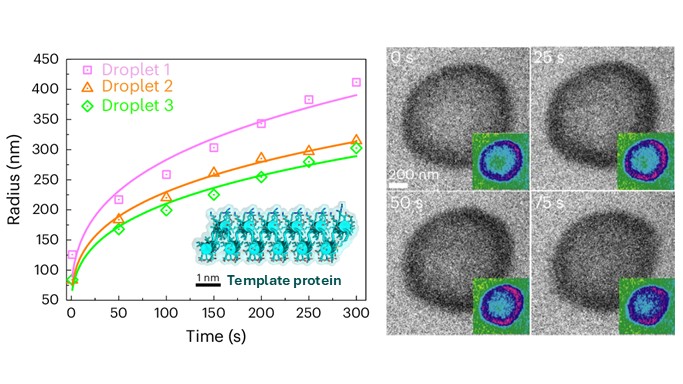
Capturing the formation and evolution of dense liquid phase of calcium (bi) carbonate
In situ liquid-phase TEM captured nonclassical calcium (bi)carbonate nucleation via a transient, highly hydrated dense liquid phase (DLP), resolving its formation and transformation in real time.
- DLPs formed in a sealed liquid cell from CaCl2, NaHCO3, and polyacrylic acid or a carboxylic acid-rich de-novo-designed helical repeat protein via liquid-liquid phase separation (LLPS) of solvated Ca2+–(HCO3-)2 complexes.
- DLP evolved from droplets to branched networks and solidified into hollow amorphous calcium carbonate.
- The findings resolve nonclassical nucleation pathways and inform control of carbonate mineralization for biomaterials, geochemistry, and CO2 sequestration.
Reference: Biao Jin, et al, Nat. Mater. 24,125–132 (2025). DOI: 10.1038/s41563-024-02025-5
Image copyright © 2024, The Author(s), under exclusive license to Springer Nature Limited

Characterization of biosensors
Observe biomolecule interactions and interfacial dynamics on biosensor surfaces in liquid environments.
Characterization of biosensors
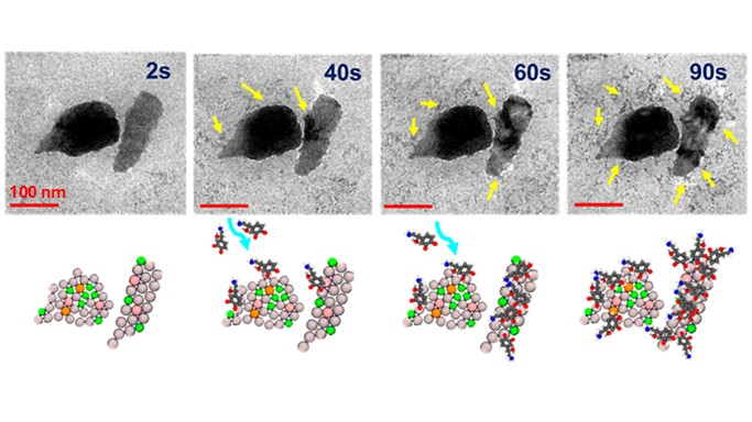
Direct visualization of biomolecule interactions at 2D materials surfaces
Interaction dynamics of norepinephrine molecules on 2D aluminum quasicrystals (Al-QCs) were imaged under liquid environment.
- Binding of norepinephrine to 2D Al-QCs imaged in real time.
- Initial binding occurred at the flake edge, then spread across the Al-QC surface.
- The findings pave the way for designing next-generation, highly selective electrochemical biosensors.
Reference: Anyesha Chakraborty, et al, ACS Appl. Mater. Interfaces 17, 68552−68565 (2025) DOI: 10.1021/acsami.5c10972
Image copyright © 2025 American Chemical Society

Metal-organic frameworks (MoFs) dynamics
Investigate the structural dynamics and phase behavior of metal-organic frameworks during in situ heating.
Metal-organic frameworks (MoFs) dynamics
Phase transition in copper triazolate Cu(ta)2 MOFs captured via in-situ heating during microED
In-situ heating combined with microED was used for direct atomic-scale tracking of structural changes in individual Cu(ta)2 MOF nanocrystals.
- Cu(ta)2 nanocrystals were heated from 25 °C to 200 °C inside the XtaLAB Synergy-ED, enabling full 3D diffraction (±75° tilt).
- The α → β transition was captured in the same crystal, consistent with prior PXRD/SCXRD studies, and yielding the first single-crystal structure of the β-phase.
- This approach enables direct correlation of thermal stimuli with structural response, opening new opportunities to study phase behavior in MOFs and related porous materials.
Reference: Lee Daniels, et al, Struct. Dyn. 12, A235 (2025). DOI: 10.1063/4.0000541
Image from Structural Dynamics under the Creative Commons license

Cells and proteins in liquids
Investigate the structure, dynamics, and functional behavior of biological systems in liquid environments.
Cells and proteins in liquids
In-situ liquid phase imaging and electron holography of magnetite nanocrystals inside bacterial cells
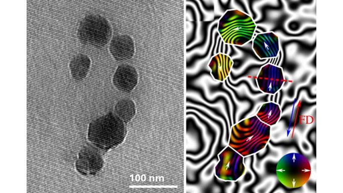
Intact bacterial cells were imaged in liquid while mapping magnetic fields from intracellular magnetite nanocrystals using off-axis electron holography, enabling simultaneous structural and electromagnetic characterization.
- Bacterial cells were encapsulated in a ~200-800 nm thick liquid layer.
- In-situ magnetization was achieved by tilting the specimen ±75° and using magnetic field of the objective lens.
- Magnetic induction maps of magnetite chains were reconstructed in liquid, yielding saturation magnetization values (~0.58–0.63 T).
- This work highlights in situ liquid-phase electron holography for probing electromagnetic behavior in biological systems, enabling studies of nanoparticle interactions, biomineralization, and protein electrostatics.
Reference: Tanya Prozorov, et al, J. R. Soc. Interface 14, 135, 20170464 (2017). DOI: 10.1098/rsif.2017.0464
Image copyright © 2017 The Royal Society Publishing

Microgel formation and growth
Study the growth, organization, and structural dynamics of microgels and hybrid soft materials in liquid environments.
Microgel formation and growth
Tracking nanoscale structural dynamics of microgel-nanoparticle hybrid systems in aqueous environments
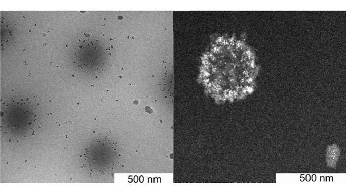
Synthesis of a hybrid microgel system of poly(N-vinylcaprolactam) (PVCL), glycidyl methacrylate (GMA), and FePt nanoparticles (NPs) was imaged using in-situ liquid phase TEM.
- Aqueous dispersion of PVCL/GMA microgels were loaded with sub-10 nm FePt NPs in a liquid cell followed by HAADF-STEM imaging.
- FePt NPs were distributed throughout the microgels, with a concentration gradient from the shell toward the core and localized along microgel fingers.
- Higher loadings led to non-uniform distribution and smaller nanoparticle clusters.
- The results highlight the utility of liquid phase TEM for studying nanoscale organization in hydrated soft materials under complex liquid environments.
Reference: K. Wiemer, et al, J. Mater. Chem. B 5, 1284-1292 (2017). DOI: 10.1039/C6TB02342H
Image copyright © 2017 Royal Society of Chemistry

Micelle formation and dynamics
Observe micelle formation, nanoparticle encapsulation, and self-assembly dynamics in liquid environments.
Micelle formation and dynamics
Dynamic micellization and gold nanoparticle encapsulation in block copolymers revealed via in-situ liquid phase imaging
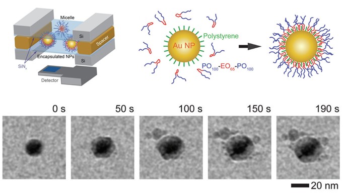
Micelle growth in aqueous (ethylene oxide)100-block-(propylene oxide)65-block-(ethylene oxide)100(EO100-PO65-EO100) block copolymer solution was imaged with and without gold nanoparticles (NPs).
- EO100-PO65-EO100 solution above critical micelle concentration (CMC) was used for tracking micelle growth.
- Polystyrene-capped gold NPs were introduced to visualize encapsulation dynamics within the polymer system.
- Micelles nucleated and grew into core–corona structures; encapsulation of hydrophobic nanoparticle occurred through polymer adsorption.
- These insights guide the design of nanocarriers and future in situ studies of controlled NP loading and release processes.
Reference: Chang Li, et al, Nanoscale 11, 2299-2305 (2019). DOI: 10.1039/C8NR08922A
Image copyright © 2019 Royal Society of Chemistry

Crystal orientation mapping
Investigate crystal orientation and structural organization of biominerals using correlative three-dimensional characterization.
Crystal orientation mapping
Crystal orientation mapping of coral skeleton microstructures
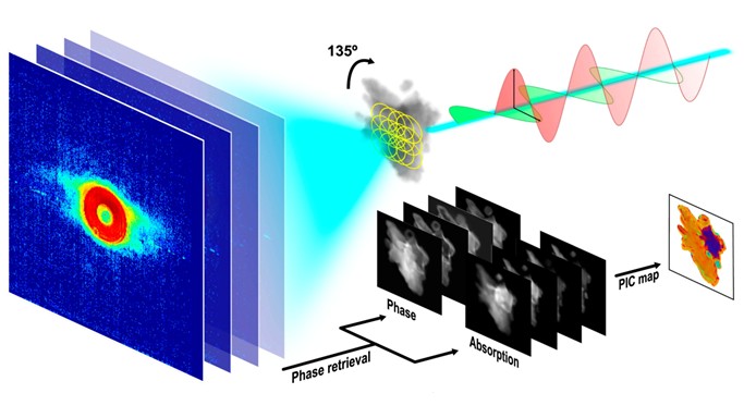
X-ray linear dichroic ptychography and 4D-STEM were used to map the crystal orientation of CaCO3 nanocrystals in coral skeletons.
- Ptychography phase and adsorption images revealed the presence of differently oriented nanograins across the coral fragment down to 35 nm resolution.
- The findings were confirmed by 4D-STEM and electron diffraction analyses.
- The study showcases a correlative workflow for X-ray and electron microscopy for studying structure and formation of biominerals.
Reference: Yuan Hung Lo, et al, Proc. Natl. Acad.Sci. 118, 3, e2019068118 (2021) DOI: 10.1073/pnas.2019068118
Image copyright © 2021 National Academy of Sciences
Biomaterial induced corrosion
Visualize biomaterial-induced corrosion processes and nanoscale structural evolution in real time under liquid environments.
Biomolecule-templated growth
Capture biomolecule-templated mineral growth and self-assembly while directly observing nanoscale structural evolution.
Biomineralization processes
Visualize nucleation, growth, and phase evolution during biomineralization under liquid-phase conditions.
Characterization of biosensors
Observe biomolecule interactions and interfacial dynamics on biosensor surfaces in liquid environments.
-dynamics_thumbnail.jpg)
Metal-organic frameworks (MoFs) dynamics
Investigate the structural dynamics and phase behavior of metal-organic frameworks during in situ heating.
Cells and proteins in liquids
Investigate the structure, dynamics, and functional behavior of biological systems in liquid environments.
Microgel formation and growth
Study the growth, organization, and structural dynamics of microgels and hybrid soft materials in liquid environments.
Micelle formation and dynamics
Observe micelle formation, nanoparticle encapsulation, and self-assembly dynamics in liquid environments.
Crystal orientation mapping
Investigate crystal orientation and structural organization of biominerals using correlative three-dimensional characterization.
Browse publications
Journal of Hazardous Materials
CrystEngComm
ACS Nano
Nature Materials
Research Spotlight
Video showing growth and dissolution of amorphous CaCO3 observed in the presence of sodium polyacrylate under liquid cell TEM.
Biomineral growth and dissolution under liquid environments
Understanding how biominerals form through shape-preserving transformations—from transient amorphous precursors to crystalline phases—unlocks new routes to engineer complex architectures beyond traditional crystallographic limits. The Hummingbird Scientific liquid flow sample holder enables direct, real-time visualization of these processes across diverse liquid environments, revealing the nanoscale mechanisms that drive structure formation.
The video captures the dynamic growth and dissolution of amorphous CaCO₃ in the presence of sodium polyacrylate (PAA), revealing real-time cycles of formation, dissolution, and re-nucleation. These observations uncover distinct transformation pathways governed by additive interactions. Notably, crystalline CaCO₃ emerges during the dissolution of ACC, highlighting a coupled dissolution–recrystallization mechanism that directs phase evolution.
Hummingbird advantages
- Stable imaging under continuous liquid flow enabling uninterrupted observation of dynamic processes.
- Nanoscale visualization revealing transformation pathways and phase evolution in real time.
Reference: Zhaoming Liu, et al, Proc. Natl. Acad. Sci. (2021) DOI: 10.1073/pnas.1914813117
Video Copyright © 2020 National Academy of Sciences
Why choose Hummingbird Scientific for your research?
Hummingbird Scientific supports in situ bio/soft materials research with flexible microscopy platforms, customizable sample chips, direct scientist support, internal TEM lab expertise, and in-house engineering and manufacturing capabilities.
Broader options for in situ bio/soft materials experiments
Bio/soft materials research spans biomineralization, biomolecule interactions, protein self-assembly, hydrogels, polymers, biosensors, metal-organic frameworks (MOFs), hydrated biological specimens, and other dynamic soft matter systems. Hummingbird supports these workflows with TEM, SEM, and X-ray microscopy platforms for continuous liquid flow, optional liquid mixing, heating, electrical biasing, and optical illumination. This gives researchers more ways to match the experimental platform to the biological system, sample environment, and characterization objectives.
More chip choices for experimental flexibility
Microfabricated chip design plays a critical role in bio/soft materials experiments by defining the liquid environment, sample thickness, fluid handling, and analytical performance. Through our dedicated microfabrication division, Hummingbird develops standard and custom chips for liquid flow, liquid mixing, heating, electrical biasing, spacer thicknesses, window geometries, microfluidic layouts, and specialized sample architectures. This enables researchers to tailor the experimental platform to their biological or soft material system while maintaining compatibility across TEM, SEM, and synchrotron X-ray microscopy.
More chip choices for experimental flexibility
Bio/soft materials researchers work directly with scientists and technical staff who understand the practical challenges of imaging hydrated, beam-sensitive, and dynamic specimens. Hummingbird's internal TEM Lab allows our team to evaluate microscope-facing performance during development, including sample handling, liquid flow, imaging stability, environmental control, and workflow usability under real experimental conditions.
Engineering, production, and custom capability
Hummingbird's in-house capabilities connect engineering, microfabrication, manufacturing, assembly, calibration, and testing within one development process. For bio/soft materials research, that means standard products can be supported by custom chips and holder configurations, and application-specific workflow changes when the experiment requires something more specific.
Biomaterial induced corrosion
Visualize biomaterial-induced corrosion processes and nanoscale structural evolution in real time under liquid environments.
Biomaterial induced corrosion
Real-time nanoscale mapping of pentanoic acid–induced corrosion in carbon steel
In-situ imaging revealed how biofeedstock-derived acids drive corrosion in carbon steel. Correlative analysis linked microstructure to initiation and propagation.
- Liquid-phase TEM of steel lamellae in pentanoic acid, combined with TKD, EDX, and thickness mapping.
- Strain-driven corrosion observed with accelerated localized attack and minimal grain boundary activity.
- These results highlight liquid-phase TEM for probing biomaterial processing environments, enabling predictive, real-time evaluation of material stability in complex organic media.
Reference: Zhiheng Lyu, et al, ACS Nano 19, 31, 28315-28325 (2025) DOI: 10.1021/acsnano.5c06142
Image copyright © 2025 American Chemical Society
Biomolecule-templated growth
Capture biomolecule-templated mineral growth and self-assembly while directly observing nanoscale structural evolution.
Biomolecule-templated growth
Uncovering the dynamics of protein-engineered nano-calcite nucleation and assembly
De-novo helical repeat (DHR) proteins were used as programmable templates to direct controlled calcium carbonate nucleation, growth, and assembly, captured in real time via in situ liquid-phase TEM.
- DHR monomers and protein–Ca2+ assemblies were observed directly, with engineered α-helical motifs pre-organizing Ca2+ ions to mimic calcite coordination.
- Nano-calcite nucleated with non-natural {110}/{202} facets, followed by nanoparticle attachment driving oriented mesocrystal assembly with tunable size and polymorph.
- The results establish in-situ liquid phase TEM as a powerful platform for decoding and programming biomolecule-templated mineralization, enabling design of hybrid materials for carbon sequestration, catalysis, and bioinspired systems.
Reference: Fatima A. Davila-Hernandez, et al, Nat. Commun. 14, 8191 (2023). DOI: 10.1038/s41467-023-43608-1
Image copyright © 2024 Springer Nature Limited
Biomineralization processes
Visualize nucleation, growth, and phase evolution during biomineralization under liquid-phase conditions.
Biomineralization processes
Capturing the formation and evolution of dense liquid phase of calcium (bi) carbonate
In situ liquid-phase TEM captured nonclassical calcium (bi)carbonate nucleation via a transient, highly hydrated dense liquid phase (DLP), resolving its formation and transformation in real time.
- DLPs formed in a sealed liquid cell from CaCl2, NaHCO3, and polyacrylic acid or a carboxylic acid-rich de-novo-designed helical repeat protein via liquid-liquid phase separation (LLPS) of solvated Ca2+–(HCO3-)2 complexes.
- DLP evolved from droplets to branched networks and solidified into hollow amorphous calcium carbonate.
- The findings resolve nonclassical nucleation pathways and inform control of carbonate mineralization for biomaterials, geochemistry, and CO2 sequestration.
Reference: Biao Jin, et al, Nat. Mater. 24,125–132 (2025). DOI: 10.1038/s41563-024-02025-5
Image copyright © 2024, The Author(s), under exclusive license to Springer Nature Limited
Characterization of biosensors
Observe biomolecule interactions and interfacial dynamics on biosensor surfaces in liquid environments.
Characterization of biosensors
Direct visualization of biomolecule interactions at 2D materials surfaces
Interaction dynamics of norepinephrine molecules on 2D aluminum quasicrystals (Al-QCs) were imaged under liquid environment.
- Binding of norepinephrine to 2D Al-QCs imaged in real time.
- Initial binding occurred at the flake edge, then spread across the Al-QC surface.
- The findings pave the way for designing next-generation, highly selective electrochemical biosensors.
Reference: Anyesha Chakraborty, et al, ACS Appl. Mater. Interfaces 17, 68552−68565 (2025) DOI: 10.1021/acsami.5c10972
Image copyright © 2025 American Chemical Society
Metal-organic frameworks (MoFs) dynamics
Investigate the structural dynamics and phase behavior of metal-organic frameworks during in situ heating.
Metal-organic frameworks (MoFs) dynamics
Phase transition in copper triazolate Cu(ta)2 MOFs captured via in-situ heating during microED
In-situ heating combined with microED was used for direct atomic-scale tracking of structural changes in individual Cu(ta)2 MOF nanocrystals.
- Cu(ta)2 nanocrystals were heated from 25 °C to 200 °C inside the XtaLAB Synergy-ED, enabling full 3D diffraction (±75° tilt).
- The α → β transition was captured in the same crystal, consistent with prior PXRD/SCXRD studies, and yielding the first single-crystal structure of the β-phase.
- This approach enables direct correlation of thermal stimuli with structural response, opening new opportunities to study phase behavior in MOFs and related porous materials.
Reference: Lee Daniels, et al, Struct. Dyn. 12, A235 (2025). DOI: 10.1063/4.0000541
Image from Structural Dynamics under the Creative Commons license
Cells and proteins in liquids
Investigate the structure, dynamics, and functional behavior of biological systems in liquid environments.
Cells and proteins in liquids
In-situ liquid phase imaging and electron holography of magnetite nanocrystals inside bacterial cells
Intact bacterial cells were imaged in liquid while mapping magnetic fields from intracellular magnetite nanocrystals using off-axis electron holography, enabling simultaneous structural and electromagnetic characterization.
- Bacterial cells were encapsulated in a ~200-800 nm thick liquid layer.
- In-situ magnetization was achieved by tilting the specimen ±75° and using magnetic field of the objective lens.
- Magnetic induction maps of magnetite chains were reconstructed in liquid, yielding saturation magnetization values (~0.58–0.63 T).
- This work highlights in situ liquid-phase electron holography for probing electromagnetic behavior in biological systems, enabling studies of nanoparticle interactions, biomineralization, and protein electrostatics.
Reference: Tanya Prozorov, et al, J. R. Soc. Interface 14, 135, 20170464 (2017). DOI: 10.1098/rsif.2017.0464
Image copyright © 2017 The Royal Society Publishing
Microgel formation and growth
Study the growth, organization, and structural dynamics of microgels and hybrid soft materials in liquid environments.
Microgel formation and growth
Tracking nanoscale structural dynamics of microgel-nanoparticle hybrid systems in aqueous environments
Synthesis of a hybrid microgel system of poly(N-vinylcaprolactam) (PVCL), glycidyl methacrylate (GMA), and FePt nanoparticles (NPs) was imaged using in-situ liquid phase TEM.
- Aqueous dispersion of PVCL/GMA microgels were loaded with sub-10 nm FePt NPs in a liquid cell followed by HAADF-STEM imaging.
- FePt NPs were distributed throughout the microgels, with a concentration gradient from the shell toward the core and localized along microgel fingers.
- Higher loadings led to non-uniform distribution and smaller nanoparticle clusters.
- The results highlight the utility of liquid phase TEM for studying nanoscale organization in hydrated soft materials under complex liquid environments.
Reference: K. Wiemer, et al, J. Mater. Chem. B 5, 1284-1292 (2017). DOI: 10.1039/C6TB02342H
Image copyright © 2017 Royal Society of Chemistry
Micelle formation and dynamics
Observe micelle formation, nanoparticle encapsulation, and self-assembly dynamics in liquid environments.
Micelle formation and dynamics
Dynamic micellization and gold nanoparticle encapsulation in block copolymers revealed via in-situ liquid phase imaging
Micelle growth in aqueous (ethylene oxide)100-block-(propylene oxide)65-block-(ethylene oxide)100(EO100-PO65-EO100) block copolymer solution was imaged with and without gold nanoparticles (NPs).
- EO100-PO65-EO100 solution above critical micelle concentration (CMC) was used for tracking micelle growth.
- Polystyrene-capped gold NPs were introduced to visualize encapsulation dynamics within the polymer system.
- Micelles nucleated and grew into core–corona structures; encapsulation of hydrophobic nanoparticle occurred through polymer adsorption.
- These insights guide the design of nanocarriers and future in situ studies of controlled NP loading and release processes.
Reference: Chang Li, et al, Nanoscale 11, 2299-2305 (2019). DOI: 10.1039/C8NR08922A
Image copyright © 2019 Royal Society of Chemistry
Crystal orientation mapping
Investigate crystal orientation and structural organization of biominerals using correlative three-dimensional characterization.
Crystal orientation mapping
Crystal orientation mapping of coral skeleton microstructures
X-ray linear dichroic ptychography and 4D-STEM were used to map the crystal orientation of CaCO3 nanocrystals in coral skeletons.
- Ptychography phase and adsorption images revealed the presence of differently oriented nanograins across the coral fragment down to 35 nm resolution.
- The findings were confirmed by 4D-STEM and electron diffraction analyses.
- The study showcases a correlative workflow for X-ray and electron microscopy for studying structure and formation of biominerals.
Reference: Yuan Hung Lo, et al, Proc. Natl. Acad.Sci. 118, 3, e2019068118 (2021) DOI: 10.1073/pnas.2019068118
Image copyright © 2021 National Academy of Sciences
Featured products


Frequently asked questions
Ready to discuss your experiment?
Talk with an applications scientist to identify the right products for your experiment and request a quote.