A History of Electron Microscopy Software

From digital imaging and analytical microscopy to open, automated, and AI-ready scientific workflows.
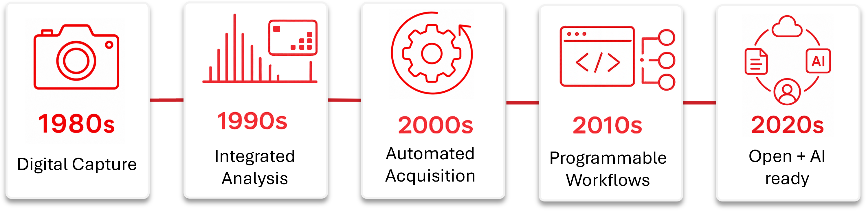
Electron microscopy software has evolved from separate tools for capturing images and spectra into increasingly connected environments for operating microscopes, coordinating detectors and stages, automating acquisition, analyzing large datasets, and preserving experimental context.
This evolution reflects a broader change in electron microscopy. Software no longer supports only the observation and analysis of a sample. It increasingly helps coordinate the complete experiment, including microscope operation, detector acquisition, specimen position, environmental conditions, data processing, and decisions about what should happen next.
1980s: The beginning of digital electron microscopy workflows
During the 1980s, most electron microscopes still relied on analog controls, photographic film, phosphor screens, and dedicated detector electronics. Software was available, but it was usually connected to individual instruments or analytical systems rather than the complete microscope.
Early software supported digital image capture, EDS and EELS acquisition, image digitization, basic processing and measurement, diffraction analysis, and instrument diagnostics.
These tools established the first major transition in electron microscopy: moving from analog observation toward digitally captured and processed scientific data.
1990s: Digital cameras, analytical integration, and spectrum imaging
The 1990s brought wider use of digital cameras, software-controlled spectroscopy, and electronic data storage. Researchers increasingly expected images, spectra, diffraction patterns, and related settings to be collected and reviewed digitally.
Platforms such as Gatan DigitalMicrograph, Emispec ES Vision, analySIS, and iTEM combined camera acquisition, image analysis, spectroscopy, measurement, reporting, and data management.
Electron tomography also began creating new software requirements. Tilt-series acquisition, alignment, and reconstruction required closer coordination between the microscope, stage, camera, and analysis workflow.
Software was beginning to move beyond individual measurements and toward more integrated experiments.
2000s: Microscope control, tomography, scripting, and automated acquisition
During the 2000s, software began controlling more of the microscope and acquisition process. Microscope optics, scanning, stage movement, cameras, detectors, spectroscopy, and acquisition sequences became increasingly connected.
Platforms such as FEI TIA supported integrated TEM, STEM, EDS, and EELS workflows. Commercial and academic tomography tools coordinated tilt-series acquisition, alignment, reconstruction, and visualization.
SerialEM advanced automated tomography, montaging, calibration, and microscope scripting. Leginon brought automated navigation, target selection, imaging, and decision-making into cryo-EM workflows.
By the end of the decade, electron microscopy software was becoming a coordinated environment for acquisition, analysis, and automation.
2010s: Direct detectors, cryo-EM, 4D-STEM, Python, and workflow software
The 2010s brought direct electron detectors, faster cameras, automated cryo-EM, 4D-STEM, in situ microscopy, and rapidly growing data volumes.
Software such as EPU, Velox, DigitalMicrograph, SerialEM, and PyJEM supported more automated, programmable, and multimodal workflows. Python interfaces also made it easier to connect microscope operation with custom scripts, external hardware, and scientific computing.
SEM and FIB-SEM software also became more programmable, supporting automated imaging, stage navigation, site selection, patterning, analytical workflows, and correlative experiments.
Open-source tools such as ImageJ, Fiji, HyperSpy, LiberTEM, py4DSTEM, Jupyter, and napari expanded electron microscopy beyond the microscope room and into reproducible analysis, high-performance computing, visualization, and machine learning.
The emphasis was shifting from operating the instrument to managing the complete workflow.
2020s: Open ecosystems, connected instruments, and increasingly autonomous experiments
The 2020s are being shaped by a new question: how should microscopes, detectors, stages, sample environments, automation tools, AI systems, and data infrastructure work together?
Current software environments increasingly support remote operation, automated acquisition, large multidimensional datasets, Python integration, AI-assisted target selection, connected analysis, and shared facility workflows.
Open-source tools continue to expand alongside OEM platforms such as Velox, Smart EPU, PyJEM, and FEMTUS. The result is not one universal software system, but a growing ecosystem of connected tools.
Electron microscopy software is moving from controlling individual instruments toward coordinating complete scientific experiments.
What this history points toward
The next stage of electron microscopy software will require more than faster acquisition or better image analysis.
Future workflows may need to observe a sample, interpret what is changing, select the next measurement, adjust position or experimental conditions, verify the result, and preserve the context behind every action.
That requires reliable control, open and documented interfaces, synchronized experimental data, persistent sample identity, accurate stage coordinates, calibration, defined safety boundaries, and human oversight.
The future of electron microscopy will depend on cooperation between microscope manufacturers, detector and hardware developers, research facilities, open-source communities, institutional systems, and customer-developed software.
Related Colibri Platform™ Pages
Explore how Colibri supports open, structured, and portable in-situ TEM data workflows:
- Colibri Platform™
- Hummingbird Control™ Software
- Hummingbird Connect™ OEM Integration
- In-Situ Experiment Data Management
- LIMS and Institutional Data
- AI Ready TEM, STEM, and SEM Workflows
- The Future of Open Microscopy